Movement:
In order for humans to move about the world, they first need muscle cells (Suny). Muscle cells are specialized for contractility, and can be classified according to their microscopic appearance (Suny). They can also be sorted into three major categories including smooth muscle, skeletal muscle and cardiac muscle (Suny). Smooth muscle is usually found in tubular organs, where as skeletal muscle is usually attached to bones (Suny). Lastly, cardiac muscle is usually found in the wall of the heart (Suny).
In order for humans to move about the world, they first need muscle cells (Suny). Muscle cells are specialized for contractility, and can be classified according to their microscopic appearance (Suny). They can also be sorted into three major categories including smooth muscle, skeletal muscle and cardiac muscle (Suny). Smooth muscle is usually found in tubular organs, where as skeletal muscle is usually attached to bones (Suny). Lastly, cardiac muscle is usually found in the wall of the heart (Suny).

http://www.cytochemistry.net/microanatomy/muscle/smooth1.jpg
The smooth muscle cell is spindle-shaped, and has an elongated nucleus that is in the middle of the cell (Suny). Smooth muscle cells may occur as solitary fibers such as those found in the spleen capsule, or grouped together in bundles known as fascicles (Suny). These fascicles can be isolated, or can be in sheets around tubular organs or vessels (Suny). Blood vessels can also be found between these fascicles (Suny).
There are also the skeletal muscle, that are unlike the smooth muscle cell, and do not have one nucleus, but many nuclei (Suny). This nuclei is also not located in the center of the cell, but rather the periphery of the cell, located just under the sarcolemma (Suny). The fiber of this cell is also filled with parallel myofibrils, which extend the length of the cell (Suny). The cross-banding of the adjacent myofibrils are then aligned with each other so the banding is able to be seen across the whole fiber (Suny).

Lastly, the cardiac muscle found around the heart, are equally important (Suny). The longitudinal sections of cardiac fibers, make it so the myofibrils can be seen by the branching of the fibers (Suny). Like the smooth muscle, the nuclei is located centrally. Also like the skeletal muscle, there are several nuclei, not just one as in the smooth muscle (Suny).

http://www.besthealth.com/besthealth/bodyguide/reftext/images/100085.jpg
Muscle cells, contraction and calcium:
In order for humans to move and use their limbs for complex activities, we must first look at muscle contraction. Muscle contraction occurs when a muscles cell lengthens or shortens (Mader). The process of locomotion, found in more complex animals, is possible only through the repeated contraction of many muscles at certain times (Mader). Contraction is actually controlled by the central nervous system, which is comprised of mostly the brain and spinal cord (Mader). The brain is what controls voluntary muscles contractions, while the spinal cord controls involuntary reflexes (Mader).
 http://www.bodytrends.com/articles/strength/images/musclefail_100x136.jpg
The term contraction actually implies that something is being shortened or reduced in the muscular system, and refers to the generation of force by muscle fibers (Mader). During muscular contraction, a muscle’s length can decrease, increase or remain constant (Mader). There are two main types of contraction of muscles: concentric, and eccentric (Mader). Concentric contraction is a a type of muscle contraction where the muscles shorten while generating force (Mader). During this type of contraction, a muscle is stimulated to contract according to the sliding filament mechanism (Mader). This actually occurs throughout the length of the muscle, which generates force at the musculo-tendinous junction (Mader). This causes the muscle to shorten and change the angle of the joint, making us able to move (Mader). If we used the elbow joint as an example, a concentric contraction of the biceps would cause the arm to bend at the elbow, and the hand to move from the leg close to the shoulder, known as a bicep curl (Mader).
In eccentric contraction, the force opposing the contraction of the muscle is actually greater than the force produced by the muscle (Mader). Instead of working to pull a joint in the direction of the muscle contraction, the muscle slows the movement of a joint, and lengthens while generating force (Mader). Even though this force is generated by the muscle is less than the force opposing contraction, it may still be below the maximal force to muscle the muscle could potentially produce (Mader). Again, using the example of the elbow joint, in relation to the arm, an eccentric contraction of the biceps muscle, the elbow would straighten and a hand would move from the shoulder to thigh (Mader). Muscles are more likely to undergo heavy eccentric loading is it is suffering from greater damage (Mader). In other words, if you do too much exercise, you will exert an overload of strain on your body, which makes your muscles soar and tired (Mader).
Movement across joints:
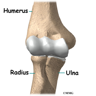
When it comes to joints, most of them can be considered freely movable joints (Frolich). The joints consist of the joint capsule, articular cartilage, synovial membrane, and synovial (joint) cavity (Mader). There aer also six classification of freely movable joints: ball in-socket, condyloid, gliding, hinge, pivot, and saddle (Mader). These joints are said to be more complex structures than immovable and slightly movable joints (Mader). In these types of joints, the end of the bones are covered with a smooth layer of cartilage, and the whole joint is enclosed in a watertight membrane that contains a small amount of lubricating fluid (Mader). This lubrication is very important, because it allows the joint to work with little friction (Mader).
When it comes to joint movement, there are four main types: gliding, angular, rotation, and circumduction. Gliding is the simplest of these motions, and moves without using any rotary or angular motion (Mader). This motion only exists between two adjacent surfaces (Mader). The next type if angular motion (Mader). Angular motion decreases or increases the angle between two adjoining bones (Mader). The most common types of angular motion are flexion, bending arm or leg, extension, straightening or unbending, abduction, and moving an extremity toward the body (Mader). The next type of joint movement is rotation (Mader). Rotation is a movement in which the bone moves around a central point without being displaces, such as turning your head from side to side (Mader).
 http://www.eorthopod.com/images/ContentImages/elbow/elbow_arthroplasty/elbow_arthro_anatomy01.jpg
Bones, bone tissue, and calcium:
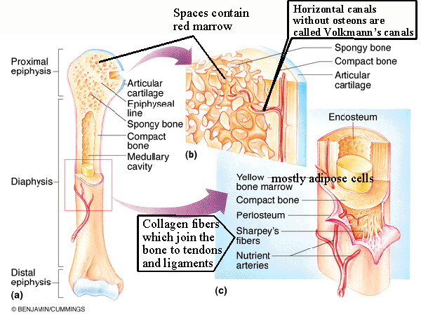
Bone tissue, also known as osseous tissue, is the major structural and supportive connective tissue of the body (Mader). Osseous tissue forms in the rigid part of the bone organs which make up the skeletal system (Mader). This type of bone tissue is a mineralized connective tissue (Mader). Bone-forming cells called osteoblasts deposit a matrix of collagen, but also release calcium, magnesium, and phosphate ions (Mader). All of these things chemically combine and harden within the matrix (Mader). This combination of hard mineral and flexible collagen makes bone harder than cartilage without being brittle (Mader).
There are two main types of osseous tisse: compact, and spongy (Mader). Compact bone forms the extremely hard exterior while spongy bonds fills the hollow interior (Mader). The tissues are biologically identical, and the difference is in how the microstructure is arranged (Mader).
Osseous tissue is important is performing numerous functions such as support for muscles, organs, and soft tissues (Mader). This tissue is also used for leverage and movement, and used to protect vital organs such as the heart (Mader). Osseous tissue is also important for calcium phosphate storage (Mader).
 http://www.unm.edu/~jimmy/long_bone.jpg
Bones are different than bone tissue (Mader). Bones are organs that are made up of bone tissue, as well as marrow, blood vessels, epithelium, and nerves (Mader). Bone tissues is only the mineral matrix that form the rigid sections of the organ (Mader).
Citations:
Frolich, Larry. “Nervous Function Powerpoint” pg. 3-7
Mader, Robert. “Human Biology 10th ed”. 2008.
Suny Downstate Medical Center. “Muscle” http://ect.downstate.edu/courseware/histomanual/muscle.html
1 comment:
Tamara, great, practically perfect work on this unit. I appreciated all the different aspects of your model limb and your compendium reviews are very complete. The leech lab should have had screen shots of the sensory neuron that you were able to impale and get data from, but just a minor thing. Keep up the hard work—just one unit to go!
LF
Post a Comment